Cranial cruciate dog ligament rupture: why does it tear?
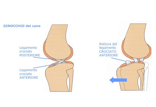
The Cranial cruciate dog ligament is the most important structure in stabilizing the dog’s knee. It’s a ligament structure, 30mm long and 11mm wide inside the joint. It has two very important jobs:
. Prevents over-extension of the knee
. Limits the rotation of the tibia
What is the Cranial cruciate dog ligament Rupture (CCLR)?
We have a rupture when a ligament structure (or part of it) is torn. A case of CCLR must undergo a surgery, since simple medicaments won’t fully heal the patient. As proof of that, a treatment that involves only anti-inflammatories or likewise will only delay the problem without never fully healing.
The cranial cruciate dog ligament Rupture can be partial or complete
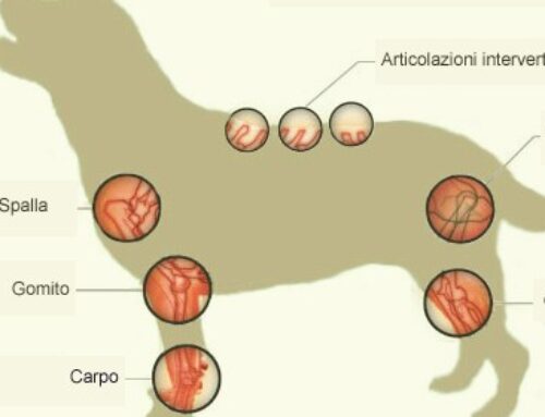
CCLR is a very common disease, found in every breed of dog, with bigger, heavier and/or more active patients at a slightly higher risk. The wound has various degrees of knee damage and pain, especially during the days immediately after, with a chance of arthrosis in the most chronic cases.
Why does the Cranial cruciate dog ligament rupture?
The ligament will rupture because of a morphological predisposition of the tibia in some breeds.
While in the human the same ligament wound has usually a traumatic origin, in the dog the reasons are rarely the same, since they usually are:
. Overweight
. Obesity
. Metabolic diseases
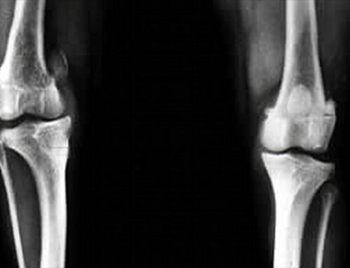
Straight and angular limbs
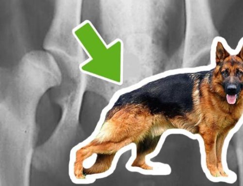
In some breeds with very straight limbs (Bulldogs and Chow Chow, for example), the tibial plate is quite inclined. This implies a greater stress on the ligament every time the dog is standing, which will lead to wearing and, in time, breaking. Other breeds like the German Sheperd have the back limbs more angular, which means the risk a rupture is lower. On a side note, even smaller dog can suffer from CCLR, often caused by a pre-existing condition.
What are the symptoms of Cranial cruciate dog ligament rupture?
CCLR is often displayed by a lameness of the back limbs.
He/she shows pain when sitting and, in most case, won’t be able to sit well.
Temporary improvement?
– After the first days, it may seem that the lameness is gone, but in reality, it’s just your dog avoiding to use his limbs properly to avoid the pain. Without treatment, the knee will suffer from an inflammation and the lameness will return stronger than before.
Untreated partial ruptures will become complete
Using the limb while broken, will create damages to joint structures like the meniscus.
The meniscus, if wounded, will bring to a worsening of the problem. In the case of a wounded meniscus, you could hear a specific sound during the exam of the knee, called “meniscus clank”. The best tip is to keep the dog resting as to avoid further damage to the joints.
How to diagnose a cranial cruciate dog ligament rupture?
To get a proper diagnosis, it’s vital to observe the dog walking from the very first moment he/she enters the room. The doctor will examine the knee the same way humans do, then will check the swelling of the knee area before taking the x-ray
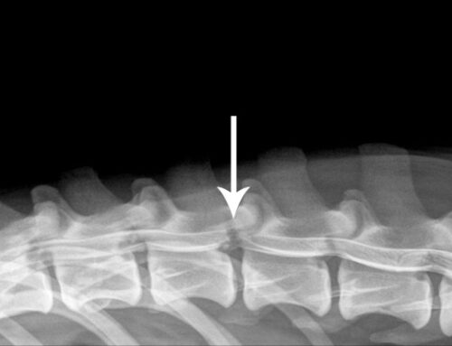
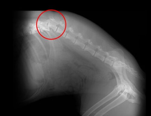
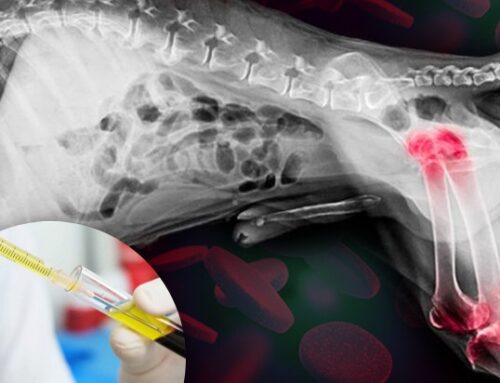
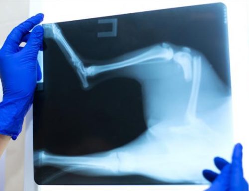
X-ray and synovial fluid
The x-ray exam is performed while the patient is sedated, and is needed to fully understand the tibial plate inclination, the arthrosis degree and all the damages derived from a rupture. With some patients, a synovial fluid analysis is needed, to cross out eventual infections or other diseases.
What are the solutions to cranial cruciate dog ligament rupture?
After a diagnosis, the solution is always surgical. The target of the operation is to stabilize the knee, eliminate the pain and diminish the development of arthrosis. The usual first step is to perform a little arthrotomy to check the meniscus damage and evaluate how to act from there.
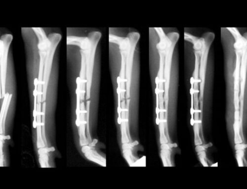
What operation: Traditional or Biomechanical?
There are two kinds of surgery: the traditional kind, which aims at the stabilization of the wounded ligament, and the biomechanical, that modifies the structure of the knee in order to make the cranial cruciate ligament not needed.
Traditional operation
Formed by intracapsular and extracapsular techniques, its purpose is to stabilize the knee by using textures or similar solutions, putting them in the same place of the wounded ligament. It is mostly used for adult patients under 15kg with a sedentary life. As a side note, it is possible to operate cats with the same procedure.
Biomechanical operation
– There are lots of biomechanical operations and the most common is the Proximal Tibial Osteotomy. It has been researched for over 20 years and it allows for an optimal recovery of the limb’s functions. It consists in rotating the tibia, thus reducing the inclination of the tibial plate. The stabilization is given by a plaque made for this kind of operations. Full recovery of the limbs occurs in the following four months, when the dogs can return to their normal activities, sports included.